Urinary System
The urinary system removes waste products from the blood and excretes urine. It also regulates the chemical and water balance of the blood.
Osmoregulation and Excretion
To survive, all animals need to maintain concentrations between water and solutes in their body within a certain range.
They can maintain this balance in two ways. One is to be an osmoconformer and have body fluids with solute concentrations equal to their surroundings. Most marine invertebrates are osmoconformers, balancing concentrations in relation to seawater. The second is to be an osmoregulator and have your internal solute concentrations independent from the outside environment. Freshwater and land-dwelling animals and many marine invertebrates are also osmoregulators. This control over concentrations of solutes in cells to prevent excess uptake or loss of water is known as osmoregulation.
Osmoregulation creates waste and waste disposal is an important aspect of this process as most metabolic waste is dissolved in water to be removed from the body. Metabolism produces a number of toxic by-products and through excretion, the process where animals dispose of metabolic wastes, they are eliminated.
Types of Metabolic Waste Products
The type of waste products produced and how they are excreted depends on the animal’s evolutionary history and habitat.
Most aquatic animals dispose of their nitrogenous waste as ammonia (NH3). It is too toxic to be stored in the body, but it is very soluble and diffuses rapidly across cell membranes. If an animal is surrounded by water, ammonia readily diffuses out of its cells and body. Small soft-bodied invertebrates like jellyfish excrete ammonia while fishes excrete it through thin membranes on their gills.
Because it is so toxic, ammonia can be tolerated at very low concentrations and must be transported in dilute solutions. Since most terrestrial animals cannot afford to lose water in amounts needed to routinely excrete ammonia, they convert it into less toxic compounds that can be safely stored in the body. The disadvantage, though is that energy is needed for this to occur.

Mammals, adult amphibians, sharks, and some bony fishes excrete urea as their major waste product. It is a soluble form of nitrogenous waste that is produced in the liver by a metabolic cycle that combines ammonia with carbon dioxide. An advantage to excreting urea is its very low toxicity.
Some animals can switch between excreting ammonia and urea depending on environmental conditions. Some toads excrete ammonia as tadpoles but excrete urea as land-dwelling adults.
Insects, land snails, and many reptiles, including birds, convert ammonia to uric acid to avoid water loss completely. Uric acid is a water-insoluble precipitate and so water isn’t used to dilute it. It is a relatively nontoxic nitrogenous waste that can be safely transported and stored in the body to be released periodically by the urinary system.
In most cases, uric acid is excreted as a semisolid paste (the white material we see in bird droppings is mostly uric acid). Although an animal must expend more energy to excrete uric acid, this is balanced by great savings in body water.
The Human Urinary System
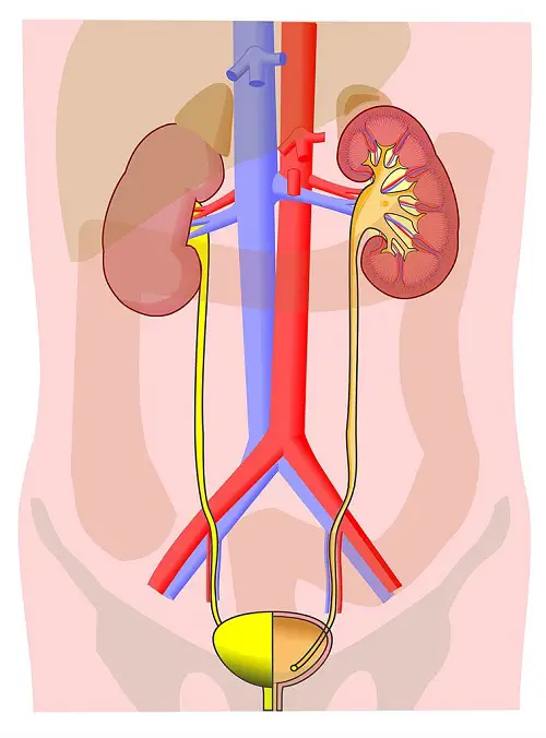
The urinary system plays an important role in homeostasis, forming and excreting urine while regulating the amount of water and solutes in body fluids. The main processing centers of this system in our body are our kidneys, compact organs located towards the back of the abdomen.
The kidneys are made of small tubes, called tubules, providing an extensive area for exchanging solutes, water, and wastes. Associated with the tubules is an intricate network of blood capillaries.
The human body contains about five liters of blood that circulates repeatedly and so, about one to two thousand liters pass through the kidneys every day. From the blood, our kidneys extract 180 liters of fluid, called filtrate, which are composed of water and other valuable nutrients that must be kept and recycled by the body in order to prevent losing nutrients and dehydration.
In a process known as filtration, the pressure of the blood forces water and other small molecules through a capillary wall into a kidney tubule. The final product of this process is urine which leaves each kidney through a duct called a ureter.
Both ureters drain to the urinary bladder and during urination, urine is expelled from the bladder through a tube known as the urethra which empties either through an orifice near the vagina in females or through the penis in males. A sphincter near the bladder and urethra controls the flow of urine.

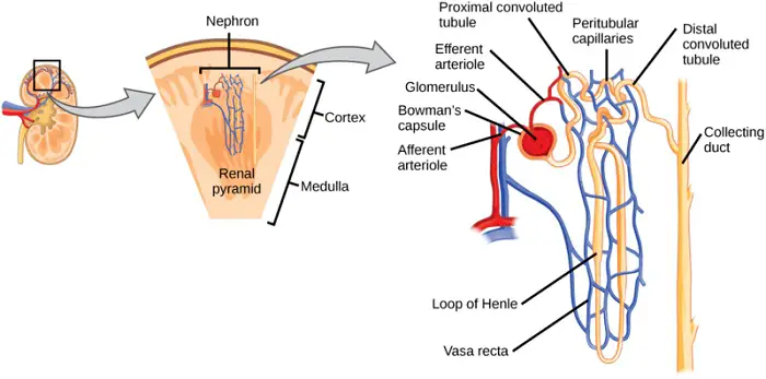
The kidney has two main regions: an outer renal cortex and an inner renal medulla. Functional units called nephrons compose the kidney. These nephrons work by extracting a tiny amount of filtrate from the blood and processing it into a smaller quantity of urine.
A nephron is made of a folded tubule and associated blood vessels which highlight the association between blood vessels and the nephron in refining the filtrate. The blood-filtering end of the nephron is a cup-shaped swelling known as the Bowman’s capsule located in the kidney’s cortex. Nephrons extend to the medulla then loop back to the cortex, meeting the collecting duct which carries urine through the medulla into the renal pelvis.

Blood enters the nephron from a branch of the arterial blood vessel (renal artery) and flows into a ball of capillaries called a glomerulus. The glomerulus and the surrounding Bowman’s capsule make up the blood filtration unit of the nephron. Filtration here leaves blood cells and large molecules such as plasma proteins behind in the capillaries.
The filtrate forced into the Bowman’s capsule flows into the nephron tubule where it is processed in three major sections of the nephron: (1) the proximal tubule; (2) the loop of Henle, a hairpin loop with a network of capillaries; and (3) the distal tubule which drains it into a collecting duct and so on until it exits the body.
The importance of having the nephrons associated with the capillaries is because of reabsorption, the process by which water and important solutes are reclaimed from the filtrate by cells making up the tubules and returned to the blood.
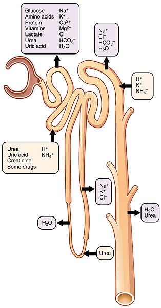
In contrast, some substances are transported from the blood into the filtrate in a process called secretion. Examples are when there are high H+ ions that are secreted into the filtrate as they affect blood pH levels. Finally, in excretion, urine is passed from the kidney towards the outside of the body.
Reabsorption is a complex and important function of our urinary system. A closer look at the process will tell us how the structures of the kidney and input from other organs facilitate the process.
A Closer Look at Reabsorption
The kidneys are able to reabsorb almost 99% of the filtrate we produce each day. This efficient water reabsorption is related to the anatomy of the nephron and the pathway of the filtrate through the kidneys. Water is not actively pumped across a membrane; it flows passively. Our kidneys reclaim water from the filtrate by moving solutes; the water then follows by osmosis and facilitated diffusion.
Most of the reabsorption of glucose, amino acids, and other valuable solutes such as NaCl occurs in the proximal tubules. As these solutes are transported, water is followed by osmosis. About 65% of the water in the filtrate is reabsorbed in the proximal tubules. As the filtrate makes its way through the rest of the nephron and collecting duct, a gradient in the concentration of solutes allows further reabsorption of water from the filtrate into the interstitial fluid.
In the long loop of Henle, the filtrate is carried deep into the medulla then back to the cortex. Water exits the filtrate as it flows down the loop of Henle due to solute concentrations of the filtrate being lower than the interstitial fluid in the medulla. Aquaporins facilitate the movement of water out of this region of the nephron. Removed from the filtrate, the water moves quickly into nearby blood capillaries otherwise if left in the medulla it will destroy the gradient needed for reabsorption.

Water reabsorption stops just after the filtrate rounds the hairpin turn in the loop of Henle; this is because this part of the organ lacks aquaporins. Although water isn’t exiting the filtrate as it travels from the medulla back to the cortex, NaCl is moving out. This movement of NaCl is the main driver of high solute concentration in the interstitial fluid of the medulla.
In the distal tubule, water exits the filtrate by osmosis while NaCl and other molecules are reabsorbed from the filtrate. Final processing of the filtrate occurs as the collecting duct carries the filtrate through the medulla. In the medulla, some urea leaks into the interstitial fluid, increasing solute concentrations further and helping maintain the solute gradient.
As the filtrate moves through the collecting duct, more water is reabsorbed before the final product, urine is passed into the renal pelvis.
It is mentioned throughout this system that maintaining a proper concentration of solutes in the body is important. Consider then what happens when we lose water by sweating. As our bodies become dehydrated, the solute concentration rises and when it gets too high, receptors in the brain signal the pituitary gland to secrete the hormone called antidiuretic hormone (ADH) into the blood.
This hormone binds to receptor molecules on epithelial cells in the collecting ducts of the kidney, leading to a temporary increase in aquaporins in the plasma membrane of the cells. This will lead to greater reabsorption of water and decrease the amount of water excreted (resulting in concentrated, darker urine which indicates not drinking enough water).
Conversely, if too much water was drunk, the blood levels of ADH drop, decreasing the number of aquaporins. The result of this would be clearer and watery urine. Diuresis is increased urination (because ADH acts against it, it is called the antidiuretic hormone) and diuretics, such as alcohol, are substances that inhibit the release of ADH, resulting in excessive urinary water loss. This effect contributes to dehydration and symptoms of a hangover when drinking alcohol. Coffee is also a diuretic, and so are tea and cola.
A person can survive with one functioning kidney but if both fail, the buildup of toxic wastes and lack of osmoregulation can be fatal. Fortunately, some functions of the kidneys can be performed artificially through a medical treatment called dialysis.
Our kidney’s regulatory functions are controlled by an elaborate system of checks and balances that include other hormones besides ADH. We will look into these hormones and the organ system responsible for regulating them next.
Next topic: Endocrine System
Previous topic: Lymphatic System
Return to the main article: Animal Form and Functions (Overview)
Download Article in PDF Format
Test Yourself!
1. Practice Questions [PDF Download]
2. Answer Key [PDF Download]
Copyright Notice
All materials contained on this site are protected by the Republic of the Philippines copyright law and may not be reproduced, distributed, transmitted, displayed, published, or broadcast without the prior written permission of filipiknow.net or in the case of third party materials, the owner of that content. You may not alter or remove any trademark, copyright, or other notice from copies of the content. Be warned that we have already reported and helped terminate several websites and YouTube channels for blatantly stealing our content. If you wish to use filipiknow.net content for commercial purposes, such as for content syndication, etc., please contact us at legal(at)filipiknow(dot)net