Musculoskeletal System
Also known as the locomotor system, the musculoskeletal system is composed of two broad systems that together provide the body with stability, support, shape, and movement.
The skeletal system supports the body and protects the organs. It provides a framework for muscle movement. On the other hand, the muscular system moves the body, maintains posture, and produces heat. Learn more about the musculoskeletal system in this concise reviewer.
Movement and Locomotion
Movement is a distinct characteristic of animals with most animals being fully mobile.
Locomotion, the active travel from place to place, requires energy. Major modes of animal locomotion include swimming, walking, running, hopping, crawling, and flying. All these modes must be able to counteract the forces that keep animals stationary: friction and gravity.
In order to resist the forces of friction and gravity, the body of an animal must be properly supported. A skeleton has many functions, including this support. There are three main types of skeletons found in animals: hydrostatic skeletons, exoskeletons, and endoskeletons.
Hydrostatic skeletons consist of fluid held under pressure to maintain a body; this is quite different from what we imagine skeletons are which are made of harder materials. Earthworms are examples of animals with these skeletons.
Exoskeletons are rigid, external skeletons that we found mostly in arthropods, such as crabs. An exoskeleton is composed of nonliving material so it does not grow, hence why we see insects molt when they develop.
An endoskeleton is found within the body.
The Vertebrate Skeleton

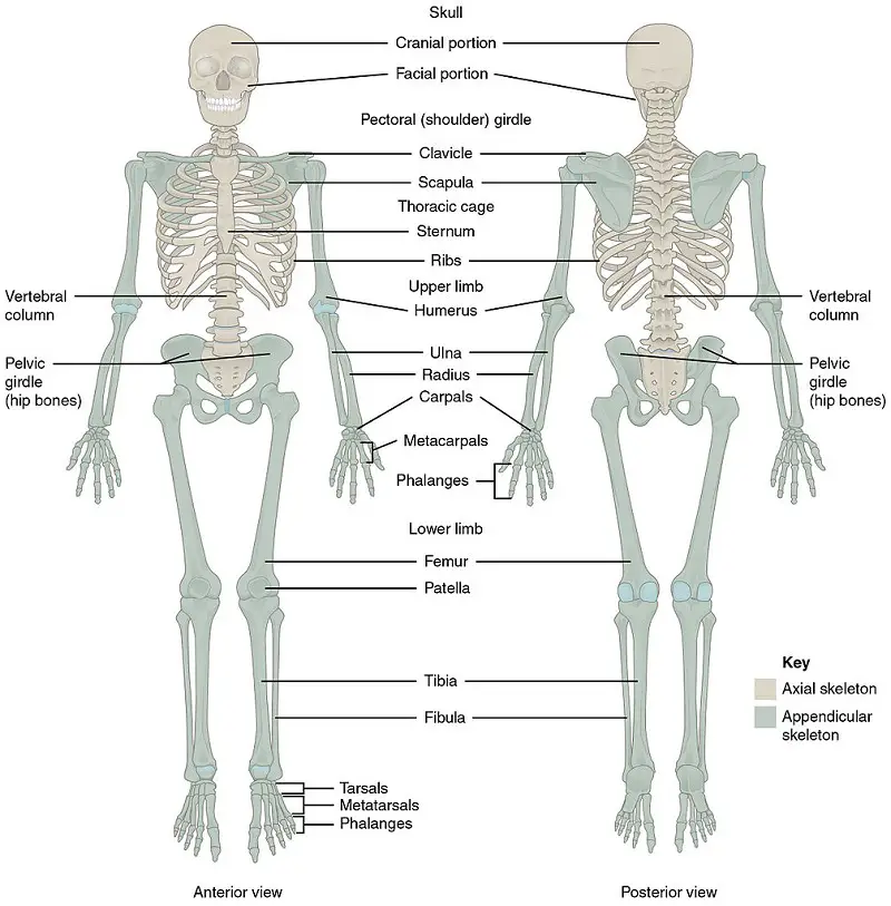
All vertebrates have an axial skeleton consisting of the skull, the backbone, and the rib cage. Attached to this is the appendicular skeleton which anchors appendages to the axial skeleton.
A bone makes up the skeleton. Compact, dense bones surround a central cavity. This central cavity contains yellow bone marrow, where fat from the blood is stored. The ends of a bone have an outer compact bone and an inner layer of spongy bone, the latter named because of its many small cavities which contain the red bone marrow that produces our blood cells.
Much of the versatility of the skeleton comes from the diverse joints. Bands of strong fibrous connective tissue called ligaments hold together the bones of movable joints. Ball-and-socket joints are joints that allow us to rotate our arms and legs. Hinge joints allow for movement in a single direction such as those in our elbows and knees. Lastly, a pivot joint allows us to rotate the forearm at the elbow.
We have focused on the skeletal system first, now we will look into how muscles function in tandem with our bones.
Muscle Contraction and Movement
Muscles are connected to bones by tendons. When a muscle contracts or shortens, it pulls along the bone to which it is attached, leading to movement. A different muscle functions to reverse the action. This back-and-forth movement is acted by muscles that come in pairs.
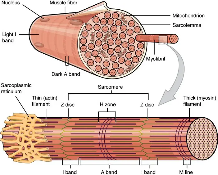
A muscle is made of many bundles of muscle fibers. Muscle fibers are long, cylindrical cells with many nuclei. Most of its volume is occupied by numerous myofibrils, bundles of proteins that include actin and myosin. Skeletal muscles are striated due to a repeating pattern of stripes along the myofibril, called a sarcomere.
A sarcomere is located between two dark, narrow lines, called Z lines, in the myofibril. Alternating bands of primarily actin and myosin create a horizontal pattern. These bands are referred to as thin filaments and thick filaments, respectively.

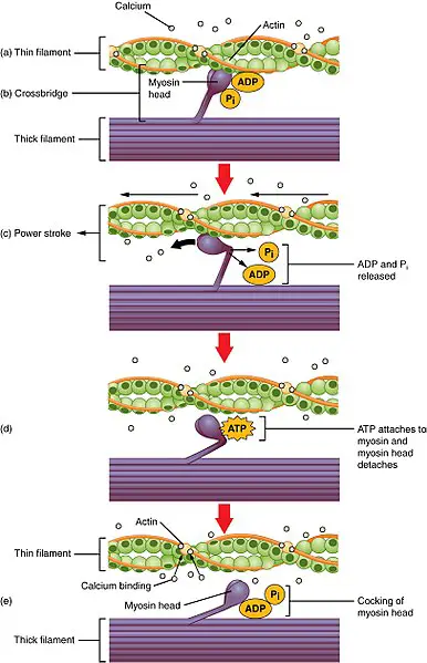
The structure of a sarcomere relates to its function: when a sarcomere contracts, its thin filaments slide along its thick filaments which is how muscle contraction fundamentally performs. Contraction shortens the sarcomere without changing the lengths of thick and thin filaments with myosin acting as the engine of the movements.
Each myosin molecule has a “tail” region and a globular “head” region. The tails are on the thick filament while the heads stick out to the side. The head has two binding sites that match the actin molecule, found in the thin filament. When ATP binds to actin, the energy powers muscle contraction by means of pivoting the head (called a power stroke) such that it will pull the thin filament towards the center of the sarcomere.

If ATP is always present, do our muscles always contract? No, this is because signals from the CNS are conveyed by motor neurons in order to initiate and sustain muscle contraction. Otherwise, we would all just be convulsing all the time.
We have tackled the different organ systems in animals. In the next topic, we will examine plant tissues and the different organ systems they comprise.
Next topic: Plants (Forms and Functions)
Previous topic: Nervous System
Return to the main article: Animal Form and Functions (Overview)
Download Article in PDF Format
Test Yourself!
1. Practice Questions [PDF Download]
2. Answer Key [PDF Download]
Copyright Notice
All materials contained on this site are protected by the Republic of the Philippines copyright law and may not be reproduced, distributed, transmitted, displayed, published, or broadcast without the prior written permission of filipiknow.net or in the case of third party materials, the owner of that content. You may not alter or remove any trademark, copyright, or other notice from copies of the content. Be warned that we have already reported and helped terminate several websites and YouTube channels for blatantly stealing our content. If you wish to use filipiknow.net content for commercial purposes, such as for content syndication, etc., please contact us at legal(at)filipiknow(dot)net
