Nervous System
The nervous system coordinates body activities by detecting stimuli, integrating information, and directing responses. Learn more about this intricate organ system with the help of this reviewer.
Nervous System: An Overview
Previously, we mentioned how the endocrine system is responsible for sending lower, more sustained responses. By contrast, the nervous system, as an organ system responsible for coordinating body functions, enables rapid coordination.
Communication in the nervous system relies on neurons, nerve cells that transmit information through electrical and chemical signals. It consists of a cell body and long thin extensions that convey signals.
With a few exceptions, the nervous system has two main anatomical divisions. The central nervous system (CNS), composed of the brain and, in vertebrates, the spinal cord; and the peripheral nervous system (PNS) which contains all the other neurons that carry information in and out of the CNS. When neurons of the PNS are bundled together and wrapped in connective tissue, they form nerves.
In addition, the PNS has clusters of neuron cell bodies called ganglia found alongside the spinal cord or those near or within the organs they serve. The PNS handles the thousands of incoming and outgoing signals that pass through the nerves and serve all areas of the body.
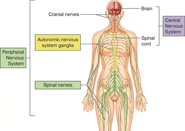
The nervous system has three interconnected functions:
- Sensory input – signals from sensory receptors that flow through the PNS to the CNS.
- Integration (in the brain and spinal cord) – interprets the signals to form appropriate responses.
- Motor output – is produced when the signals from integration enter the PNS to effector cells that will perform the response.
Meanwhile, three types of neurons correspond to the nervous system’s aforementioned three main functions:
- Sensory neurons – convey signals from sensory receptors to the CNS.
- Interneurons – integrate data and relay the appropriate signals to other neurons.
- Motor neurons – convey signals from the CNS to effector cells. The automatic response to any stimuli produces a reflex.
Neurons perform routine tasks and have two kinds of extensions that facilitate their function: numerous dendrites and a single axon. Dendrites are highly branched, often short extensions that receive signals and convey information to the cell body. In contrast, the axon is typically much longer and is responsible for transmitting signals to other cells.
The axon ends in a number of branches, each having a synaptic terminal at the end. There is a space between the terminal and another cell called a synapse, where signals are transmitted.
Neurons are only a part of the nervous system and they are supported by cells called glia. Glia may nourish neurons, insulate axons, or help maintain homeostasis of extracellular fluid surrounding neurons. An example of a glial cell is called a Schwann cell, found in the PNS. Axons that convey signals rapidly are enclosed along most of their length by a thick insulating material, kind of like the insulation used in many electrical wires, called the myelin sheath. This helps speed up the transmission of impulses along a neuron.
The gaps between Schwann cells, called nodes of Ranvier, are points on the axon where the nerve signals are to be regenerated. The said process is time-consuming. Everywhere else, the myelin sheath covers the axon and helps preserve and propagate the signal leading to a faster signal that must be regenerated constantly along the axon.
Nerve Signals and Transmission
In order to understand how nerve signals are transmitted, we will first take a closer look at a resting neuron, one that isn’t transmitting a signal.
A resting neuron has potential energy called the membrane potential, which exists as an electrical charge difference across the neuron’s plasma membrane. As a result, the inside of the cell is more negatively charged than the outside.
Because opposite charges attract, a membrane stores energy by holding opposite charges apart, like in batteries. The strength or voltage across the plasma membrane of a resting neuron is called the resting potential and it exists because of ion concentration gradients across the plasma membrane.
For most neurons, potassium ions (K+) have higher concentrations inside the cell, while sodium ions (Na+) are more numerous outside. Pumps called sodium-potassium pumps help maintain the gradient between these ions, utilizing ATP to move Na+ out of the neuron and K+ in. Ion channels are pores found throughout the membrane that help establish this resting potential.
A resting membrane has many open K+ channels and a few open Na+ channels. This will lead to potassium diffusing out the neuron more readily than sodium. And as more potassium ions move out, the inside of the cell becomes less positive, compared to its outside environment.
This electrical difference establishes the resting potential. And because the inside of the neuron becomes more negative, it attracts the positive ions back as if the internal and external environment of the neuron are playing a tug-of-war on the ions.
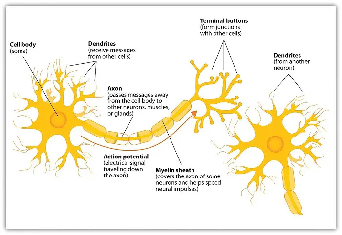
A stimulus is any factor that causes a nerve signal to be generated. Nerve signals are triggered by the use of a neuron’s potential energy. Since nerve signals can be electrical, a massive change in membrane voltage, known as the action potential, occurs as a nerve signal is transmitted along an axon. The fluctuation of an action potential is regulated by voltage-gated ion channels, structures that open and close in response to a change in membrane potential.
When an action potential reaches the end of a neuron, it is passed to a receiving cell at either an electrical or chemical synapse. In an electrical synapse, electric current flows directly from a neuron to the receiving cell through gap junctions leading to the receiving cell being stimulated quickly.
The majority of synapses are chemical and unlike electrical synapses, they have a narrow gap called a synaptic cleft which prevents the action potential from spreading directly to the receiving cell. Instead, the action potential is converted into a chemical signal in the form of neurotransmitters. This chemical signal may then generate an action potential in the receiving cell.
Some of the neurotransmitters include acetylcholine, which is released by motor neurons to activate skeletal muscles; glutamate which has a key role in forming long-term memory; and GABA (Gamma-aminobutyric acid) which acts as an inhibitory synapse in the CNS. Norepinephrine, serotonin, dopamine, and endorphins are also well-known neurotransmitters.
So far, we have introduced the nervous system and talked about nerve transmission. Now we will look at the vertebrate nervous system and this also applies to the human body.
The Vertebrate Nervous System
Structurally simpler animals, such as sponges, do not have a nervous system while other animals at the earlier branches of the evolutionary tree have a nerve net, a series of interconnected neurons throughout their body.
When animals evolved and became more complex, two changes also occurred: (1) cephalization, where the nervous system became more concentrated toward the head; and (2) centralization, the presence of a central nervous system that is distinct from the peripheral nervous system.
Vertebrates have a diversity of nervous systems but all share fundamental similarities.
1. The Central Nervous System (CNS)
The vertebrate nervous system is marked by distinct central and peripheral elements which are highly centralized. In all vertebrates, the brain and spinal cord make up the CNS while the rest of the nervous system composes the PNS.
The spinal cord is a bundle of nervous tissue that runs along the spine, conveying information between the brain and the rest of the nervous system. The master control center of the system is the brain which coordinates and regulates all actions by the organism. The circulatory system services the CNS but the animals evolved to prevent metabolic waste from entering the brain through a selective mechanism called the blood-brain barrier.
The CNS also contains fluid-filled spaces called ventricles in the brain and central canal in the spinal cord. The fluid is known as cerebrospinal fluid. Both brain and spinal cord contain white matter, made mostly of axons whose myelin sheaths are responsible for the light color, and gray matter, made mostly of neuron cell bodies and supporting glia.
2. The Peripheral Nervous System (PNS)
The PNS is made of sensory and motor nerves. Sensory neurons receive information from sensory receptors while motor neurons transmit information from the CNS to effector organs that respond to stimuli.
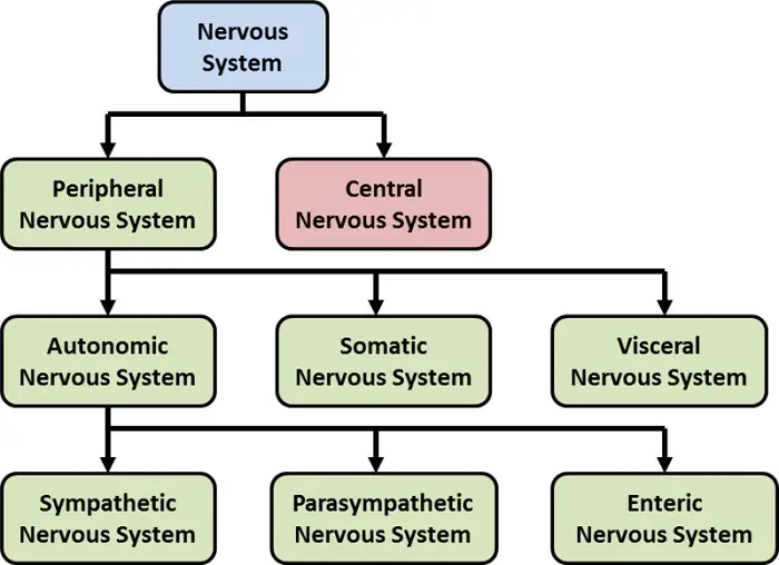
Motor neurons can be subdivided into two functional components. The motor system carries signals from the CNS to skeletal muscles and so can be voluntarily controlled. The other component is the autonomic nervous system which is generally involuntary. It is important to emphasize that both systems cooperate to maintain homeostasis.
The autonomic nervous system is composed of three divisions: parasympathetic, sympathetic, and enteric. The first two divisions have largely opposite or antagonistic effects with each other on most body organs.
The parasympathetic division prepares the body for activities that gain and conserve energy for the body (rest and digestion for example). Neurons of the sympathetic division prepare the body for energy-consuming activities.
Sympathetic and parasympathetic neurons come from different regions of the CNS and utilize different neurotransmitters. Parasympathetic neurons emerge from the lower part of the spinal cord; most use acetylcholine at synapses within target organs. In contrast, sympathetic neurons emerge from the middle regions of the spinal cord, and most release norepinephrine to target organs.
The enteric division, meanwhile, makes up neurons in the digestive tract, pancreas, and gallbladder. These neurons control secretion and peristalsis. Although it may function independently, the enteric division is normally regulated by the sympathetic and parasympathetic nervous divisions.
The Vertebrate Brain
The brain is essential for coordinating the functions of the different organ systems of the body. The structures that facilitate the functions of the brain come from three bilaterally symmetrical bulges during early embryonic development- the forebrain, midbrain, and hindbrain. The following table shows these different regions of the brain during embryonic development and the structures they will develop into:
| Brain Regions in the Embryo | Brain Structures They Form |
| Forebrain | Cerebrum Diencephalon (thalamus, hypothalamus, posterior pituitary, pineal gland) |
| Midbrain | Midbrain |
| Hindbrain | Pons Cerebellum Medulla oblongata |
An evolutionary trend observed in birds and mammals is a relatively larger brain (relative to body size) than those found in other vertebrates. This seems to explain the relatively greater capacity of birds and mammals for sophisticated behavior which correlates with the larger size of the cerebrum, the part of the forebrain responsible for cognition and integration. The cerebrum develops into two halves, the left and right cerebral hemispheres.
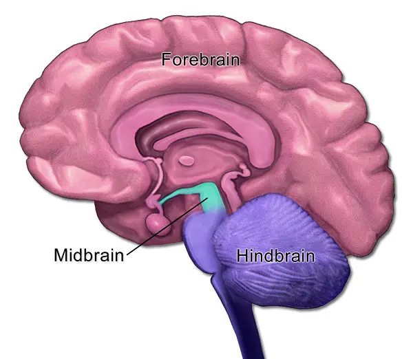
The cerebrum’s outer region, the cerebral cortex, is vital for perception, voluntary movement, and learning. In smaller mammals, this outer layer is smooth whereas in ours it is highly folded, which again means more surface area without taking up additional space.
This increase in the surface area seems to be an evolutionary advantage as it means more support for neural circuits, facilitating more complex behavior, sophisticated sensory perception, and the ability to communicate.
The Human Brain
The following table highlights the major structures of the brain and the functions they perform:
| Brain Structure | Major Functions |
| Brainstem | Conducts data to and from the brain centers Helps maintain homeostasis Coordinates large-scale body movements |
| Medulla oblongata | Controls breathing, circulation, swallowing, and digestion |
| Pons | Controls breathing |
| Midbrain | Receives and integrates auditory data Coordinates visual reflexes Sends sensory data to brain centers |
| Cerebellum | Coordinates body movement Plays a role in learning and remembering motor responses |
| Thalamus | Input center for sensory data going to the cerebrum Sorts and groups all incoming sensory data for the cerebrum |
| Hypothalamus | Homeostatic control center Controls the pituitary gland Serves as a biological clock (maintains the circadian rhythm) |
| Cerebrum | Integrates information Major role in memory, learning, speech, and emotions Formulates complex behavioral responses |
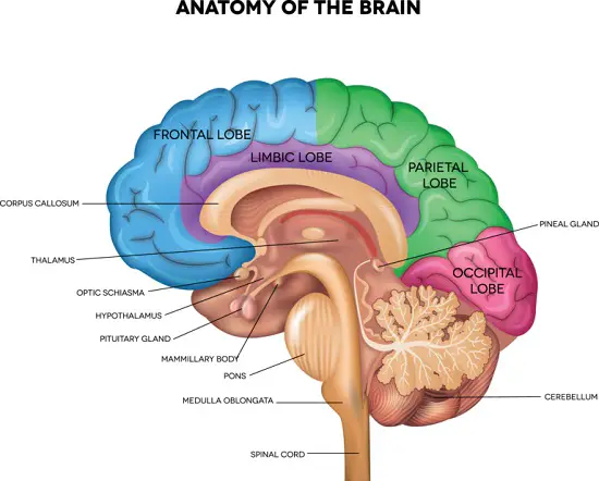
Other components of the brain that we will highlight are the left and right cerebral hemispheres of the cerebrum, each responsible for the opposite side of the body. A thick band of nerve fibers called the corpus callosum facilitates communication between the hemispheres. Under this are basal nuclei that are important in motor coordination.
You might have seen brain illustrations showing different functions. These illustrations specifically map activities to the cerebral cortex which accounts for about 80% of the total mass of the brain. This mapping of the different activities of the body to specific regions of the brain has been an ongoing task in neuroscience research. The difference in function between the right and left hemispheres is known as lateralization.
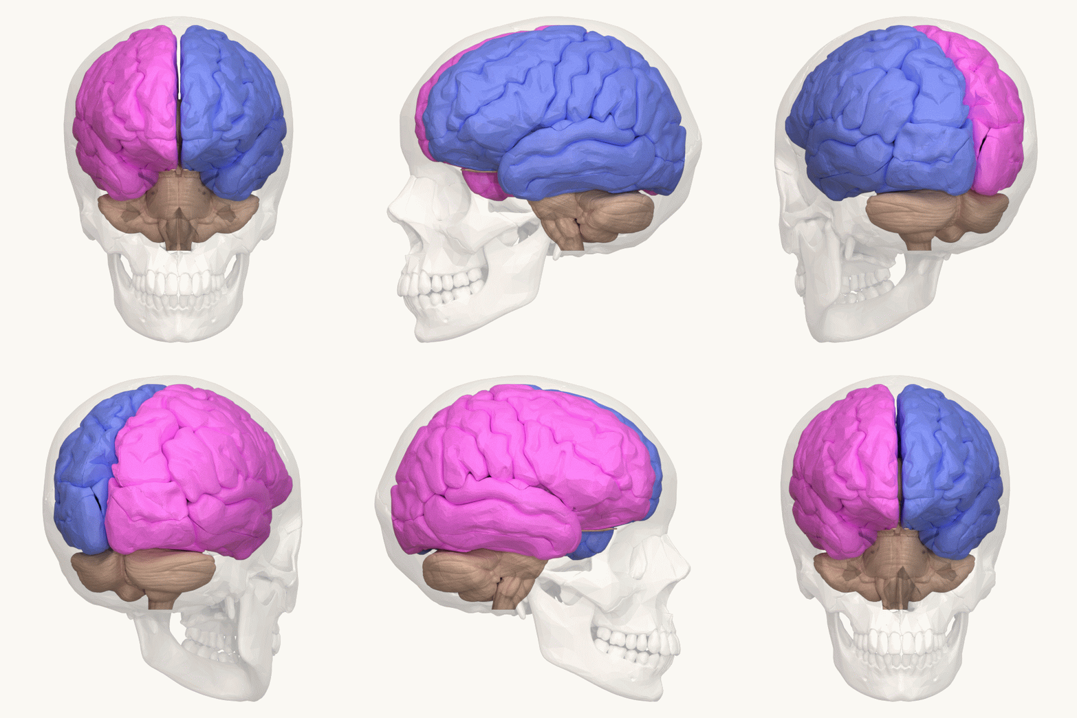
The limbic system, meanwhile, is made of different parts of the brain that function in human emotion, behavior, motivation, and memory. This functional brain system includes part of the thalamus and hypothalamus and two cerebral structures: the hippocampus, which is involved in the formation and recollection of memories, and the amygdala, which lays down emotional memories.
Changes in the brain’s physiology can lead to an enormous toll on society. Examples include depression, schizophrenia, bipolar disorder, Alzheimer’s, etc.
The brain and the nervous system facilitate all stimuli that affect the body. It is important that not only the internal body environment is regulated, but there is also awareness with regards to the external environment. Important to these are the senses, which we will tackle in the next topic.
The Sense Organs

Stimuli are detected by sensory receptors, specialized neurons (or other types of cells) that are attuned to the conditions of the external and internal environment. The reception of a stimulus leads to sensory transduction, the conversion of stimulus to a receptor potential such that it can be transmitted as a signal to the CNS.
Over time, a sensory receptor’s sensitivity to a stimulus can diminish. This phenomenon is called sensory adaptation.
Sensory receptors can be classified based on the stimulus they deal with. They are:
- Thermoreceptors detect either heat or cold.
- Mechanoreceptors respond to various forms of mechanical energy such as touch, pressure, and sound. They stimulate other mechanoreceptors to bend or stretch.
- Pain receptors respond to extreme pressure or temperature, as well as certain chemicals, all of which can damage animal tissues.
- Chemoreceptors include those in our noses and taste buds. They are attuned to chemicals in the external environment and some chemicals in the body’s internal environment.
- Electromagnetic receptors act on electricity, magnetism, or various wavelengths of light.
We will now look at the sense organs in the human body, starting with those which respond to sound and regulate our balance.
1. Hearing and Balance
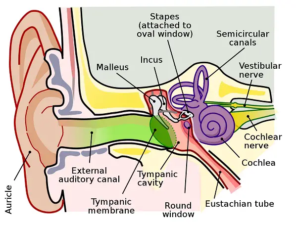
The human ear is made of two organs. One functions for hearing while the other focuses on maintaining balance. The ear has three regions: the outer ear, middle ear, and inner ear.
The outer ear consists of the fleshy flap-like structure, pinna, and auditory canal. Both collect sound waves and channel them to the eardrum, a thin sheet of tissue that separates the outer ear from the middle ear. When sound waves strike the eardrum, the vibration of the eardrum is passed on to three small bones: the hammer (malleus), anvil (incus), and stirrup (stapes). The stirrup is connected to the oval window, a membrane-covered hole in the skull through which sound is passed into the inner ear.
The middle ear opens into a passage called the Eustachian tube, connecting it to the pharynx, allowing air pressure to stay equal on either side of the eardrum (which may not vibrate properly if air pressure is unequal).
The inner ear is made of fluid-filled chambers in the bones of the skull. One of the chambers, the cochlea, is a long, coiled tube that contains the actual hearing organ, the organ of Corti, in one of its three canals. The organ of Corti is made of hair cells, embedded in a basilar membrane, which are the sensory receptors of the ear.

Lying next to the cochlea are the structures for maintaining balance: the semicircular canals, the utricle, and the saccule. The semicircular canals detect changes in the head’s rotation. Clusters of hair cells in the utricle and saccule. meanwhile, detect the orientation of the head with respect to gravity.
2. Vision
There is diversity in organs that allow animals to perceive light. Planarians, for example, have the simple light-detecting organ, the eyecup, which contains a photoreceptor cell shielded by a darkly pigmented cell.
Over the course of evolution, two types of image-forming eyes formed. The compound eye is characteristic of some invertebrates, specifically insects and crustaceans. A compound eye is made of several thousand light detectors called ommatidia. An advantage of this is how extremely sensitive they are to slight movement while in others, compound eyes provide excellent color vision. The second type of image-forming eye is the single-lens eye, which all vertebrates have.

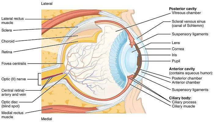
Our eyeball is surrounded by a tough, whitish layer of connective tissue called the sclera.
The eye is divided into two fluid-filled chambers: the jelly-like vitreous humor in the front chamber while the thinner aqueous humor fills the rest of the eyeball. Each humor helps maintain the shape of the eye. In addition, the aqueous humor facilitates supplying of nutrients and the removal of waste.
At the front of the eye, the sclera becomes transparent and forms the cornea, allowing light to pass through. Beneath the sclera is a pigmented layer called the choroid, the front of which forms the iris which surrounds the pupil, the opening in the center of the eye. After going through the pupil, the light passes through the lens which focuses light onto the retina where photoreceptor cells are found.
The retina is made of two photoreceptors: rods are sensitive to light and allow us to see a dim light or see white, black, and gray; and cones which are sensitive to bright light and can distinguish color. Each rod and cone has membranous disks with light-absorbing visual pigments: rhodopsin is found in rods while photopsins are found in cones (the kind of photopsin in the cone reflects the color they absorb most – red cones, blue cones, and green cones).
Three of the most common visual impairments include nearsightedness (myopia), farsightedness (hyperopia), and astigmatism. The last is caused by misshapen lenses or cornea.
Several vision problems are related to aging. Cataracts are due to the clouding of the lens while glaucoma is a group of diseases that result in damage of the optic nerve, the nerve which relays visual stimulus to the brain.
3. Taste and Smell
Our senses of smell and taste depend on receptor cells that detect chemicals in the environment. Chemoreceptors in the taste buds detect molecules in a solution while those in the nose are able to detect airborne molecules when they dissolve in the mucus that lines our nasal cavity.
Olfactory (smell) receptors line the upper nasal cavity, sending impulses along axons directly to the olfactory bulb of the brain. Olfactory receptors have cilia that contain specific receptors. And so, we are able to distinguish thousands of different odors.

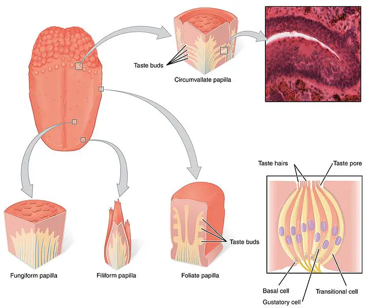
Taste receptors in mammals, meanwhile, are organized into taste buds on the tongue. Aside from the four familiar taste perceptions, a fifth one called umami is elicited by glutamate, an amino acid. Umami is the savory flavor common in meats, cheese, and other protein-rich foods.
Any region of the tongue contains all five types of taste so a “taste map” of the tongue can be misleading. The taste buds also come in different forms:
- Fungiform papillae – have a flattened head and more reddish coloration than other taste buds due to receiving a greater blood supply. They are associated with the taste of “sweet”.
- Goblet papillae – are the least abundant but most voluminous. They are responsible for sensing bitter taste and acid. They also help alert the brain if we may eat a potentially toxic substance.
- Foliate papillae – they are structurally underdeveloped but are essential in that they help us perceive the salty taste.
- Filiform papillae – though technically not taste buds as they are not directly associated with the sense of taste, they do respond to heat and touch. Keeping this in mind, they help us perceive pungent taste, which also includes “spicy”.
The senses are important in taking in information for the body to process but essential to this is how to respond as well. In the next topic, we will focus on the skeletal and muscular systems which work hand-in-hand to perform the responses.
Next topic: Musculoskeletal System
Previous topic: Reproductive System and (Embryonic Development)
Return to the main article: Animal Form and Functions (Overview)
Download Article in PDF Format
Test Yourself!
1. Practice Questions [PDF Download]
2. Answer Key [PDF Download]
Copyright Notice
All materials contained on this site are protected by the Republic of the Philippines copyright law and may not be reproduced, distributed, transmitted, displayed, published, or broadcast without the prior written permission of filipiknow.net or in the case of third party materials, the owner of that content. You may not alter or remove any trademark, copyright, or other notice from copies of the content. Be warned that we have already reported and helped terminate several websites and YouTube channels for blatantly stealing our content. If you wish to use filipiknow.net content for commercial purposes, such as for content syndication, etc., please contact us at legal(at)filipiknow(dot)net
